Magnetic Resonance Imaging (MRI)

Make an Appointment
We have locations throughout Manhattan and Westchester. View all locations and contact info.
If you have been scheduled for an MRI, it means your doctor would like to have a closer look at your tissues or organs. MRI does not use radiation.
What is MRI?
MRI uses a large magnet, radio waves, and a computer to produce detailed images of the body. An MRI exam provides doctors with a view that has greater clarity and more detail than X-ray images.
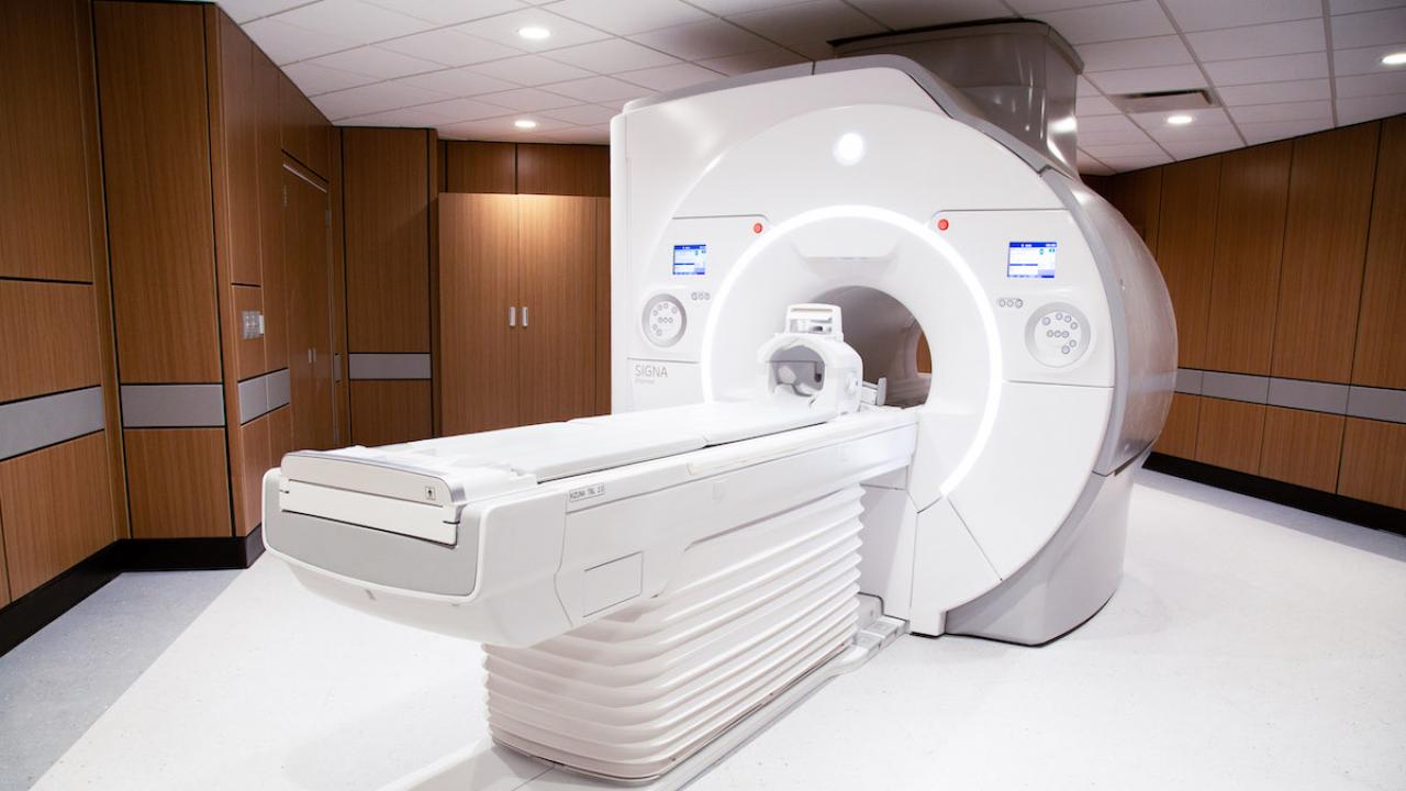
An MRI system includes a scanner shaped like a donut and a motorized table that slides in and out of the opening. The magnetic strength of the scanner is measured in Tesla, with 3 Tesla (3T) scanners providing the highest resolution images. For some exams, a lower resolution is more appropriate.
How MRI is used by doctors
Healthcare providers use MRI to evaluate nearly every part of the body and diagnose a wide range of conditions. MRI is also used to monitor response to treatments for cancer and other diseases.
Some specialized MRI exams have a slightly different preparation or procedure. You will be given any additional instructions when you schedule your exam. Specialized MRI exams include:
- Breast MRI
- Cardiac MRI
- Functional MRI (fMRI)
- Magnetic Resonance Angiography (MRA)
- MRI Enterography
- Prostate MRI
Is it safe for me to get an MRI?
For most people, an MRI is safe. You will be asked detailed questions about your medical history and any devices, implants, or other metal objects you may have in or on your body. As long as your device or implant is certified as MRI safe, you may have an MRI. If there is not enough information or your device is determined to be unsafe, your doctor will schedule a different type of imaging exam.
How do I prepare for the test?
- Eat and drink as you normally do unless you have been given other instructions when you scheduled your appointment.
- Continue to take your medications unless instructed not to.
- Talk to your doctor if you need medication for anxiety or to help you relax.
- Wear comfortable clothes to your appointment.
- Do not wear jewelry or makeup.
What will happen during the test?
- Check in for your appointment on the Connect patient portal. You may also check in when you arrive at the imaging center.
- Remove all metal objects, including jewelry, hair accessories, phones, wallets, and keys. We will give you a locker to secure them.
- We will review your medical history and MRI Safety Checklist with you.
- Some MRI exams require contrast, or dye, to help highlight certain parts of your body. If you are having an MRI with contrast, a nurse will insert an intravenous line (IV), usually in your arm. The contrast (gadolinium) will be administered during the exam.
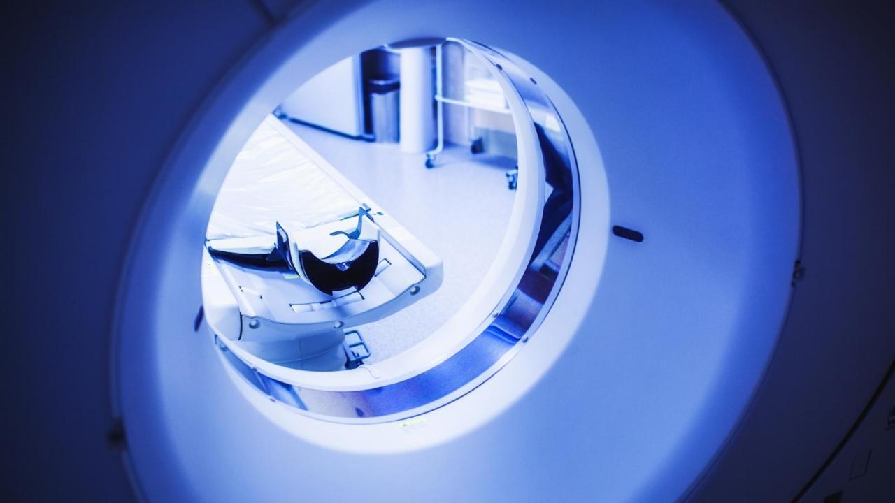
- A technologist will ask you to lie on the MRI table with your head on a pillow or in a padded cradle. The technologist may place a plastic device called a coil around the area of the body that will be looked at.
- You will be offered a blanket and earplugs. You will also be given an emergency call button in case you need to urgently contact the technologist.
- The table will slide into the scanner, and a red light may shine on your body momentarily to position the area of interest in the center of the scanner.
- The technologist will leave the room but will be able to see, hear, and speak with you at all times.
- During the scan, it is important to lie still and relax so that we can record clear images. The technologist may ask you to hold your breath for short periods of time. The scanner will make loud clicking, buzzing, banging sounds, and you may feel vibrations. This is normal and you should remain relaxed and still.
- An MRI scan can take from 15 to 90 minutes.. Most exams take between 20 and 30 minutes.
Are there any risks?
MRI does not use ionizing radiation, the type of radiation used in X-rays and CT scans. There are no known harmful side effects associated with temporary exposure to the magnetic field used by MRI scanners. There is a slight risk that you will develop an allergic reaction to the contrast (gadolinium).
After the test
After the exam you can immediately resume your normal activities. If you were given contrast, drink six to eight eight-ounce glasses of water in the 24 hours after the MRI to help remove the gadolinium dye from your body. A radiologist will analyze the MRI images and will share the results with the doctor who requested the exam. Your doctor will then discuss the results with you.