Q & A with Dr. Richard Ha: Deep Learning and Breast Cancer Risk Assessment
Breast cancer is the most common disease and second most common cause of death in women in the United States. Globally, it is number one in both categories. While mammography and other diagnostic breast imaging techniques have reduced the mortality rate of women with breast cancer over the past several decades, the question of who exactly is at risk for breast cancer remains largely unanswered.
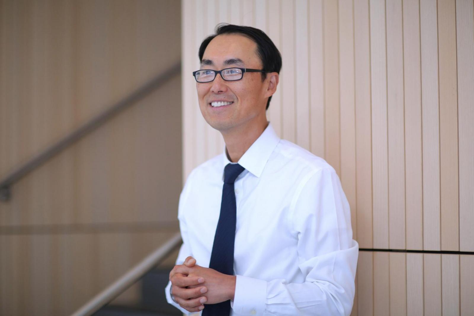
Dr. Richard Ha, Associate Professor of Radiology and the Director of Education and Research for the Division of Breast Imaging, has been applying artificial intelligence to breast imaging and breast cancer research for the past five years in an effort to answer this and other questions.
Dr. Ha and his group are using large mammographic data sets and deep learning techniques to determine women’s individual risk, with the idea that breast cancer screening guidelines could eventually become personalized—saving both lives and resources.
This Breast Cancer Awareness Month, we talked to Dr. Ha about how this and other projects his group is involved in might impact women’s health, and how his collaborations with other departments at Columbia have already shown results.
Can you start with a short overview of your research?
My area of research is in breast imaging and our research focuses on anything related to breast imaging and breast cancer, and that includes detection. Our ultimate goal is to support the clinical team, meaning we want to help our surgical colleagues and our medical oncology colleagues.
Where does artificial intelligence fit in?
One question we’re trying to answer is, what’s the best way to assess risk for breast cancer? Unfortunately, the current way of assessing risk on an individual basis is not adequate. The vast majority—about 80%—of women who get breast cancer have no family history and there are no clinical factors that we could identify that says that this person is likely to develop breast cancer. And there's not a genetic cause like BRCA in a majority of patients.
And so for this vast majority of woman who develop breast cancer, we want to see if radiology, and specifically radiomics—using deep learning—can identify features that may be predictive of an individual’s risk for breast cancer.
What exactly is radiomics?
Radiomics is a general term where you're using imaging and digitized data and you parlay that to answering clinical questions. And deep learning is part of that.
How might this research ultimately affect women?
Our research is trying to stratify women who may be at high risk versus those who may not. So if you're at a lower risk, for example, a screening guideline may look different for you—where instead of screening every year starting at age 40, perhaps you can start screening at age 45 or 50 and do it every two years. Currently, we don't have enough information either by genetics or by clinical history to really determine who should fall under the high-risk versus low-risk categories, to specifically guide screening regimen. We're hoping that by applying large data using a mammographic dataset and applying the latest technology, deep learning and artificial intelligence, we can get more information to really help distinguish the two populations.
How much do you work with other departments at Columbia?
I think we have a very powerful infrastructure here. We have excited researchers in their own discipline, and we can take their expertise and combine it with our interests to figure out how we can do the best research to impact patients.
Just to give you one example: When patients undergo a lumpectomy, which is the most common type of breast cancer surgery, we have learned from our surgical colleagues that around 20 to 40 percent of the time, patients have to come back and undergo a second surgery. And why is that? Because when they take out a specimen, they often focus around the margin to make sure that it's cancer free. Under some circumstances where there is margin positivity, a patient has to come back, go through the mental preparation again, and go through a second surgery. This is not an ideal situation.
And so we collaborated with Dr. Christine Hendon's group. She's a professor of biomedical engineering, as well as electrical engineering. Her group has developed optical coherence tomography technology, which enables us to get really high-resolution images of the specimen in real time. We collaborated with them and with our surgical colleagues and also with pathology to identify the surgical specimen image with OCT and see where the margins are positive. We've done reader studies, and then to make this really practical, we added another layer where we applied artificial neural network to interpret these images in a speedy, accurate way. And we have been very successful and in doing that.
What project are you currently working on that you’re really excited about?
We're really excited about the completion of a study that was started four years ago with our collaboration with Dr. Carolyn Westhoff's Ob/Gyn group. We set out to study if there is a way to reduce risk for breast cancer in high-risk women, with a treatment that's more tolerable. Currently there are two medications that are often used—people might've heard of Tamoxifen or aromatase inhibitor. They're effective, but they're not without side effects.
There was a company that had developed a birth control pill, called UPA, and that medication works for birth control, but our idea was to see if this could also be used to treat and reduce risk for breast cancer. We enrolled young women and we gave them a control medication as well as this birth control pill.
Our contribution to this study was, how do we assess whether this medication is working or not? And part of what's really exciting is that we now understand, based on our imaging biomarkers—the density within the breast and within MRI—that there is a specific type of normal enhancement called background parenchymal enhancement, and that's been associated with risk.
It was a double blinded study and we got great results in that it went along with our hypothesis. So, we will be enrolling and proposing a larger study to further test for efficacy. But this is just really exciting and can be potentially meaningful to many women.